
Ultrasound
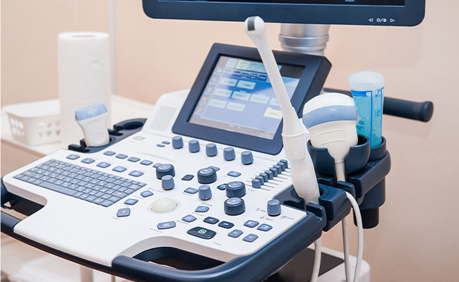
Ultrasound imaging, also known as Ultrasonography, is a method of obtaining diagnostic images from inside the human body through the use of high-frequency sound waves.
Ultrasonography is used as a diagnostic tool that can assist radiologists and technologists with making recommendations for further treatment.
 MRI
MRI CT
CT PET
PET Ultrasound
 Women's Imaging
 X-Ray
X-Ray Bone Densitometry (DEXA)
How Ultrasounds Are Used
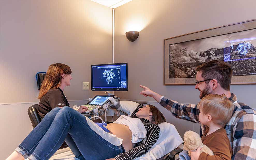
- Viewing an unborn fetus
- Examining internal organs, including the liver, gallbladder, spleen, pancreas, kidneys, bladder, uterus, and ovaries.
- Show movement of internal tissues and organs (i.e., enable radiologists to see blood flow).
- Used complementary with other procedures such as needle biopsies
- Evaluate superficial structures, such as the thyroid gland and scrotum
How to Prepare for an Ultrasound
- Wear comfortable, loose-fitting clothing.
- Depending on the type of ultrasound exam you have, you will be asked:
- Not to eat or drink for up to 12 hours before your appointment, or
- Drink up to six glasses of water two hours before your exam and avoid urinating. This will ensure a full bladder when the exam begins.
Specific Ultrasound Exam Preparations
Abdomen Ultrasound
The preparation for this exam is 8-12 hours without food or drink.
Aorta Ultrasound
The preparation for this exam is 8 hours without food or drink.
Carotid Ultrasound
There is no specific preparation for this exam.
Pelvic / Transvaginal / Bladder Ultrasound
The preparation for this exam involves having a full bladder by drinking 6-8 glasses of fluid 1-2 hours before the exam.
Renal / Renal Artery Ultrasound
The preparation for this exam is to hydrate for 6 hours before the exam and no food for 6-8 hours before the exam.
Testicular Ultrasound
There is no specific preparation for this exam.
Thyroid Ultrasound / Biopsy
There is no specific preparation for this exam.
Obstetric Ultrasound
There is no specific preparation for this exam.
Venous Leg
There is no specific preparation for this exam.
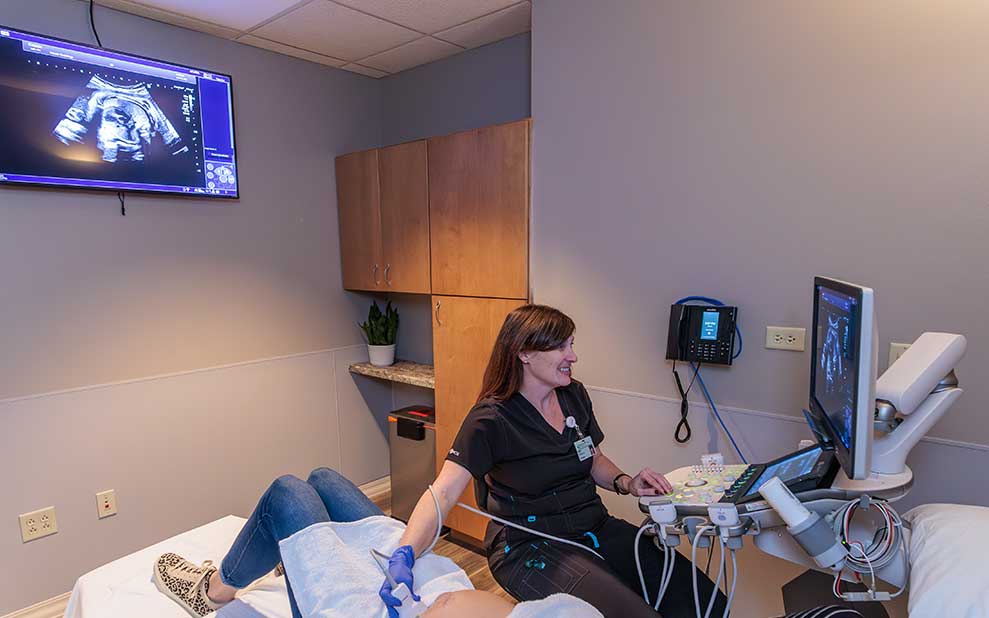
What to Expect During Your Ultrasound
- The examination usually takes less than 30 minutes.
- After being positioned on the exam table, a clear gel is applied in the area being examined. This helps the transducer make contact with the skin.
- The technologist firmly presses the transducer against the skin and moves it back and forth to image the area of interest.
- The technologist is able to review the ultrasound images in real time.
- Most ultrasound exams are painless. Gel is applied to your skin and there may be varying degrees of discomfort during the exam due to probe pressure.
Frequently Asked Questions
How long does the exam take?
The exam may take anywhere from 30 minutes - 1 hour from start to finish.
Can I eat or drink before the exam?
Depends on the exam.
Can I drive after the exam?
Yes.
Can I schedule my exam?
Imaging scans are not a self-referral exam. Your physician’s office will call us to schedule a time that is convenient for you. We will be required to have a signed order from your physician to proceed with the exam. A schedule coordinator will contact you prior to your exam to gather necessary information. This helps enable us to get you registered and back for your exam as quickly as possible.
How will I receive my results?
The specialty trained radiologists at Dakota Radiology Imaging Center will review the newly acquired images and any previous related exams. They will dictate a detailed report explaining their findings. The report is sent to your physician. Your physician will review the findings with you.
Will my insurance cover my procedure?
Many private insurance companies and Medicare provide coverage for some imaging procedures depending on medical necessity. The staff at Dakota Radiology Imaging Center.