Women’s Imaging
 MRI
MRI CT
CT PET
PET Ultrasound
 Women's Imaging
 X-Ray
X-Ray Bone Densitometry (DEXA)
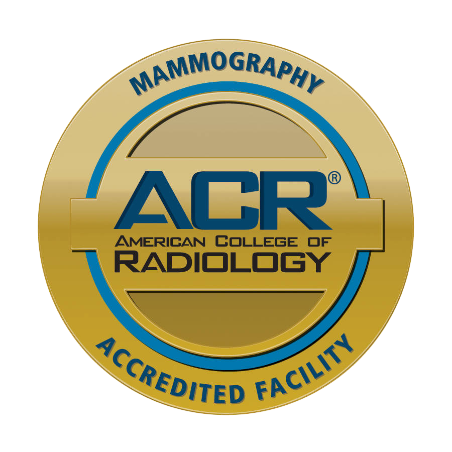
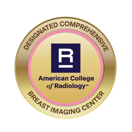
Mammography and Breast Imaging Center
Dakota Radiology offers patients and their families a comprehensive array of breast screening and women’s imaging services to provide timely information and diagnosis. To ensure the best possible experience for our patients, we offer:
- Same-day results for diagnostic patients
- A relaxing and comfortable environment
- State-of-the-art technology
Our board-certified breast imaging radiologists and technologists provide these breast imaging services:
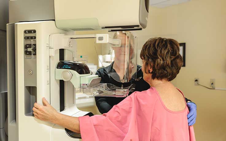
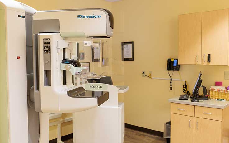
Digital Breast Tomosynthesis (3D Mammography)
Digital Breast Tomosynthesis, also known as 3D Mammography, is the latest in breast imaging technology. It creates a three-dimensional image of the breast, showing it in several ‘layers.’ By showing each layer separately, cancer is less likely to be obstructed from view by overlapping tissue.
Mammography is a critical component in the early detection of breast cancer. Screening mammography can help discover breast cancer even if there are no symptoms present. It can reveal changes in the breast before a patient or physician can identify them on an exam.
Dakota Radiology stands by the Society of Breast Imaging and American College of Radiology’s recommendations of screening mammograms beginning at age 40. We believe early detection is the best protection for finding and treating breast cancer in its early stages.
Studies have shown that 3D Mammography results in fewer callbacks and additional testing for patients. It also has shown higher cancer detection because the radiologist can view the breast in greater detail. 3D Mammography benefits all patients regardless of age, breast density, or size.
The majority of insurance providers now cover 3D, but it may vary. Please check with your provider to find out exactly what’s covered under your plan before you make your appointment.
Breast Biopsy
At Dakota Radiology, we perform three different types of breast biopsy: Ultrasound-Guided, Stereotactic, and MRI-guided. The type of biopsy performed is recommended by your radiologist for the best outcome.
Ultrasound-Guided Breast Biopsy
Ultrasound-Guided Breast Biopsy uses sound waves to help locate an abnormality or lump and remove a small tissue sample for examination. If your last breast imaging test showed an abnormal area, the radiologist might recommend doing an Ultrasound-guided biopsy of the area.
Stereotactic Breast Biopsy
Stereotactic Breast Biopsy is the latest in breast biopsy technology. New technology allows for these biopsies to be performed in the upright position while the radiologist takes a small sample of tissue to further test for breast cancer. This biopsy is done with mammographic guidance for increased accuracy and precision. A stereotactic breast biopsy exam is less invasive than a surgical biopsy and leaves little or no scarring.
MRI-Guided Breast Biopsy
An MRI-Guided Biopsy is performed to further evaluate a lesion found on a breast MRI. It is not always possible to tell from certain imaging tests if an area of concern is benign or cancerous and therefore biopsy is necessary. During an MRI-guided breast biopsy, MRI imaging is used to help the radiologist guide the biopsy needle into the site of the abnormality. A tissue sample is removed for further examination of the abnormality. Further instructions for preparation may be provided prior to your appointment. It is recommended, however, that you wear comfortable clothing and a well-fitting, supportive bra to your biopsy appointment. It is also recommended that you eat before your biopsy appointment.
Breast MRI
An MRI of the breast is an additional tool for the diagnosis of breast cancer. It is most often used in women at a high risk for breast cancer along with screening mammograms. The MRI may see cancers that a mammogram will not, but does not replace regular mammograms, as it can miss other cancers that mammogram would normally detect. A breast MRI may also be used to determine the size, location, and extent of diagnosed breast cancer. Breast MRI's require a special device called a dedicated breast coil. Not all imaging centers have this equipment, but here at Dakota Radiology, we are able to perform this type of imaging for our patients.
Seed Localizations
Seed localizations are used pre-surgery so that your surgeon can locate the seed and surrounding abnormal tissue. A tiny metal seed, smaller than a grain of rice, will be placed into abnormal breast tissue. The seed is a tiny but powerful magnet that your surgeon can later find among the abnormal tissue, especially if the abnormal tissue is too small to be seen or felt by hand. Your surgeon will then use a special tool to locate the seed within the breast and remove the seed and abnormal tissue.
Preoperative Lesion (localization before breast surgery)
After a needle breast biopsy, some lesions may require diagnostic or therapeutic surgical biopsy. This, in turn, requires accurate localization of the lesion for the breast surgeon. These localization procedures can be done under ultrasound or mammographic guidance. Patients can expect a long, thin wire to be advanced from the skin down to the area being removed. The remainder of the wire is then taped to the skin so the surgeon can follow the wire down to the lesion when in surgery.
Ductography
A Breast Ductogram is performed to evaluate nipple discharge. It is a minimally invasive and mostly painless procedure. Contrast is given during this procedure. Let your technologist or radiologist know if you have any allergies, have experienced allergic reactions to contrast in the past, or are pregnant or breast feeding.
Cyst Aspirations
A Cyst Aspiration is performed when a cyst is found through mammographic or breast ultrasound imaging and aspiration is medically necessary or recommended. Ultrasound is used for these procedures.
FRequently ASKED QUESTIONS AND INFO
What is the difference between a screening and diagnostic mammogram?
Typically, a screening mammogram is routinely administered to detect breast cancer. However, if a patient has breast pain, a lump, or an abnormal screening mammogram, they will be called back for a diagnostic mammogram to take a closer look. A low percentage of patients will require a breast biopsy.
How should I prepare for my mammogram?
Describe any symptoms you may be experiencing with your technologist before your exam. If your breasts are tender, it is recommended to take mild pain medication such as acetaminophen one hour before your exam. Do not apply deodorant, lotion or talcum powder under your arms or on your chest before your exam as they may interfere with the quality and clarity of the images.
Can I schedule my exam?
Imaging scans are not a self-referral exam. We will be required to have a signed order from your physician to proceed with diagnostic exams or procedures. A schedule coordinator will contact you to schedule your exam and to gather necessary information. This helps enable us to get you registered and back for your exam as quickly as possible.
How will I receive my results?
The specialty trained radiologists at Dakota Radiology Imaging Center will review the newly acquired images and any previous related exams. They will dictate a detailed report explaining their findings. The report is sent to your physician. Your physician will review the findings with you.
Will my insurance cover my procedure?
Many private insurance companies and Medicare provide coverage for some imaging procedures depending on medical necessity. The staff at Dakota Radiology Imaging Center will be happy to work with you to verify your benefits, eligibility and authorizations. We will submit claims as needed for your imaging scans. If the procedure cannot be covered by your insurance provider, we will be happy to develop a payment plan with you.