MRI
Magnetic Resonance Imaging, or MRI, uses a powerful field magnet and radio waves to provide clear and detailed diagnostic images of internal body organs and tissues. MRI’s are a valuable tool for the diagnosis of a broad range of conditions, including cancer, heart and vascular disease, stroke, joint and musculoskeletal disorders.
MRI differs from CT scans and X-rays, as it does not use radiation to produce images and allows the evaluation of some body structures that may not be as visible with other diagnostic imaging methods.
 MRI
MRI CT
CT PET
PET Ultrasound
 Women's Imaging
 X-Ray
X-Ray Bone Densitometry (DEXA)
Common Uses of MRI
Imaging of the Musculoskeletal System: MRI is often used to study the knee, ankle, foot, shoulder, elbow, wrist, and hand. MRI is also a highly accurate method for evaluation of soft tissue structures such as tendons and ligaments, which are seen in great detail. Even subtle injuries are easily detected. In addition, MRI is used for the diagnosis of spinal problems including disc herniation, spinal stenosis, and spinal tumors.
Imaging of the Vascular System: Magnetic Resonance Angiography (MRA) generates pictures of the arteries to evaluate them for stenosis (abnormal narrowing) or aneurysms (vessel wall dilatations, at risk of rupture). MRA is often used to evaluate the arteries of the neck and brain, thoracic and abdominal aorta, the renal arteries, and the legs (called a “run-off”). Magnetic Resonance Venography (MRV) is a similar procedure that is used to image veins.
Imaging for Cancer & Functional Disorders: Organs of the chest and abdomen such as the liver, lungs, kidney, and other abdominal organs can be examined in great detail with MRI. This aids in the diagnosis and evaluation of tumors and functional disorders. In the early diagnosis of breast cancer, MRI is an alternative to traditional x-ray mammography.
Imaging of the Brain: MRI can detect a variety of conditions of the brain such as cysts, tumors, bleeding, swelling, developmental and structural abnormalities, infections, inflammatory conditions, or problems with the blood vessels. It can detect damage to the brain caused by an injury or a stroke. MRI of the brain can also be useful in evaluating problems such as persistent headaches, dizziness, weakness, and blurry vision or seizures, and it can help to detect certain chronic disease of the nervous system, such as multiple sclerosis. In some cases, MRI can provide clear images of parts of the brain that can’t be seen as well with an X-ray, CAT scan, or ultrasound, making it particularly valuable for diagnosing problems with the pituitary gland and brain stem.
MRA
Magnetic Resonance Angiography, or MRA imaging, is an imaging test that visualizes the vascular structures of the body — more specifically, arteries and veins.
Doctors commonly order MRA’s to check for weakened or blocked arteries and vessels, to diagnose a blood clot, or to review results after vascular surgery.
How to Prepare for an MRI
- Wear loose-fitting, comfortable clothing to your exam.
- Before your MRI, let your doctor know if you have allergies, asthma, kidney or liver disease, are pregnant or might be pregnant. Your physician will let you know if you may resume or restrict any prescription medications prior to your appointment.
- Any metallic items on your body can affect the quality of the diagnostic images or may not be safe to enter the MRI suite. You will need to remove your watch, eyeglasses, jewelry, hearing aids and any other metallic objects you may have, including non-permanent dentures, prior to your exam.
- Some patients who undergo MRI in an enclosed unit may feel confined and claustrophobic. If you are not easily reassured, a sedative may be administered.
- There are no restrictions on food or drink before the exam unless instructed.
Please arrive at the to Dakota Radiology Imaging Center at least 15 minutes prior to your appointment for check-in and to fill out patient and safety forms.
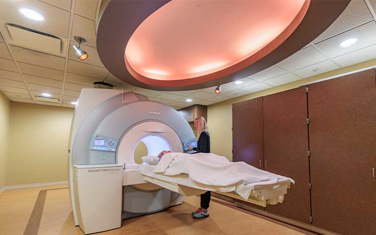
What to Expect During Your MRI
- Depending on how many images are needed, the exam generally takes 15 to 45 minutes. However, very detailed studies may take longer.
- You must lie down on a sliding table and be comfortably positioned.
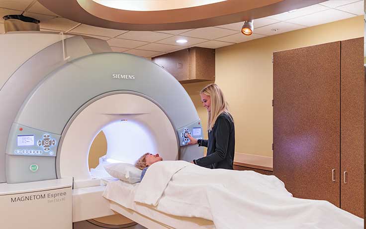
- Even though the technologist must leave the room, you will be able to communicate with them at any time using an intercom.
- If necessary, a friend or family member may be allowed to stay in the room with you during the exam.
- You will be asked to remain still during the actual imaging process. However, between sequences, which last between 2-15 minutes, slight movement is allowed.
- You will hear loud tapping or thumping noises during the exam. Earplugs or earphones may be provided to you by the MRI technologist.
- You may feel warmth in the area being examined. This is normal.
- Depending on the part of the body being examined, a contrast material may be used to enhance the visibility of certain tissues or blood vessels.
Notify your doctor or MRI technologist if you have any of the following:
- Pacemaker or implantable defibrillator
- An artificial heart valve or stent
- Body piercings
- Cochlear implants or hearing aid
- Drug pump
- Any metal dental fillings/work
- Implanted nerve stimulator
- Insulin pump
- Metal fragments, such as a bullet or shrapnel
- Metal joints or limbs
- Metal plate, pin, or other metallic implants
- Nicotine or other medication patches
- Previous gunshot wound
- Previous surgery on the area being scanned
Frequently Asked Questions
How long does the exam take?
The exam may take anywhere from 15-45 minutes from start to finish. However, very detailed studies may take longer.
Can I eat or drink before the exam?
Yes, unless instructed by your doctor or technologists.
Can I drive after the exam?
Yes, you may continue regular activities after the exam. If a prescription for claustrophobia is given, you may be required to have a ride after the exam.
Can I schedule my exam?
Imaging scans are not a self-referral exam. Your physician’s office will call us to schedule a time that is convenient for you. We will be required to have a signed order from your physician to proceed with the exam. A schedule coordinator will contact you prior to your exam to gather necessary information. This helps enable us to get you registered and back for your exam as quickly as possible.
How will I receive my results?
The specialty trained radiologists at Dakota Radiology Imaging Center will review the newly acquired images and any previous related exams. They will dictate a detailed report explaining their findings. The report is sent to your physician. Your physician will review the findings with you.
Will my insurance cover my procedure?
Many private insurance companies and Medicare provide coverage for some imaging procedures depending on medical necessity. The staff at Dakota Radiology Imaging Center will be happy to work with you to verify your benefits, eligibility and authorizations. We will submit claims as needed for your imaging scans. If the procedure cannot be covered by your insurance provider, we will be happy to develop a payment plan with you.