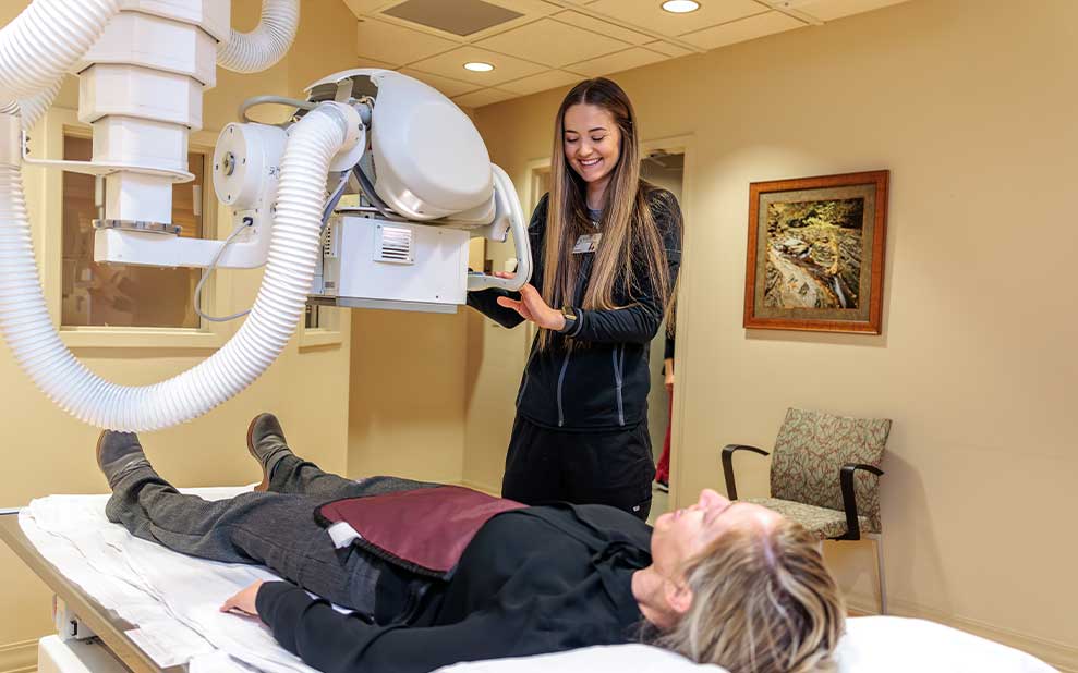
Our Imaging Services

X-ray

X-ray

X-ray is the oldest and most frequently used form of medical imaging. X-rays produce digital, diagnostic images of the human body and most commonly are used by doctors to view and assess the skeletal system for injury.

Benefits of Digital X-ray

  • Safer – less radiation than from conventional X-rays
  • Faster – takes less time because the technologist can preview your images in seconds
  • Enhanced image quality
  • More accurate

How to Prepare for an X-ray

There is no special preparation required for most X-rays. You may be asked to change into a gown prior to your examination and remove jewelry, eyeglasses and any metal objects during the exam. Women should always inform the technologist if there is any possibility that they are pregnant.

Common Uses of X-ray

  • Assist doctors in identifying and treating of bone fractures.
  • View, monitor or diagnosis joint injuries and infections, arthritis, artery blockages, abdominal pain.
  • Detection and diagnosis of cancer, although usually Computed Tomography (CT) or MRI is better at defining the extent and the nature of a suspected cancer.
  • Bone Density Scan (DXA)

What to Expect During an X-ray

  • An X-ray exam is painless and usually takes five minutes to half an hour.
  • Some discomfort may result from lying on the table, a hard surface that may feel cold.
  • Sometimes, to get a clear image of an injury such as a possible fracture, you may be asked to hold an uncomfortable position for a short time. Any movement could blur the image and make it necessary to repeat the procedure.
  • The technologist positions you on the exam table and places a cassette under the table in the area of the body to be imaged.
  • Pillows may be used to help you hold the proper position.
  • Then the technologist steps behind a radiation barrier and asks you to hold very still, without breathing for a few seconds.
  • The X-ray equipment is activated, sending a beam of X-rays through the body to expose the cassette.
  • The technologist then repositions you for another view, and the process is repeated as necessary.
  • When your X-rays are completed you will be asked to wait until the technologist checks the images.

Bone Density Scan (DXA)

A Dual-energy X-ray Absorptiometry (DXA) scan, also known as a Bone Density or Bone Densitometry Scan, is a non-invasive test that measures bone mineral density to assess if a person is at risk of fracture or osteoporosis. It is also useful in tracking the effects of treatment for osteoporosis and other conditions that can cause bone loss.

Bone density testing is recommended to the following:

  • Post-menopausal women or women age 60+ who have risk factors for developing osteoporosis
  • Patients with a personal or maternal history of hip fracture or smoking
  • Post-menopausal women who are tall (over 5 feet 7 inches) or thin (less than 125 pounds)
  • Men and women who have hyperparathyroidism
  • Men and women who have been on medications that are known to cause bone loss for an extended period

How should I prepare?

  • Refrain from taking calcium supplements for at least 24 hours beforehand.
  • Wear comfortable clothing and avoid garments that have zippers, belts or buttons made of metal.
  • Let your technologist know if you’ve recently had a barium examination, have been injected with a contrast material for a CT or MRI, or radioisotope scan.
  • Let your technologist know if there is a possibility you are pregnant.

Who interprets the Bone Density Scan results and how do I receive them?

The results of a DEXA bone density exam are interpreted by a radiologist and forwarded to your doctor. Your test results will be in the form of one of two scores:

T score – This number is used for postmenopausal women as well as men and women over the age of 50. This number shows the amount of bone you have compared to a young adult of the same gender with peak bone mass. A score above -1 is considered normal. A score between -1 and -2.5 is classified as osteopenia, the first stage of bone loss. A score below -2.5 is defined as osteoporosis. It is used to estimate your risk of developing a fracture.

Z score – This number is used for premenopausal women as well as men and women under the age of 50. This number reflects the amount of bone you have compared to other people in your age group and of the same size and gender. If it is unusually high or low, it may indicate a need for further medical tests.

Frequently Asked Questions

How long does the exam take?

The exam may take anywhere from 5 to 30 minutes from start to finish.

Can I eat or drink before the exam?

Yes, unless instructed by your doctor or technologists.

Can I drive after the exam?

Yes, you may continue normal activities after the exam.

Icon Phone WhiteSchedule your appointment today by calling (605) 721-8626